
Why a New Review on Seed Oils Misrepresents Dietary Intake, Biomarkers, and Fatty-Acid Biology
A newly published review in Current Nutrition Reports argues that linoleic acid (LA) may offer health benefits beyond cardiovascular disease, citing associations with cognition, dementia, liver fat, muscle mass, healthy aging, and skin cancer (Messina et al., 2026). The authors argue that LA has been unfairly demonized and that higher exposure could be beneficial.
But there’s a critical flaw in their reasoning.
The paper treats dietary LA intake, circulating LA, erythrocyte LA, adipose-tissue LA, and genetically predicted circulating LA as if they are interchangeable measures of the same biological exposure. They are not.
Once these distinct compartments are separated, the reassuring narrative becomes far less convincing.
The paper’s industry disclosures also warrant consideration. Main author Messina is an employee of Soy Nutrition Institute Global, which receives funding from the United Soybean Board and companies involved in the soy-food industry. Two additional authors report previous financial support from Soy Nutrition Institute Global for unrelated work.
Industry ties don’t automatically invalidate research, but they demand careful scrutiny—particularly when the exposure in question is a major component of soybean oil.
“LA Exposure” Is Not a Single Variable
The review repeatedly groups dietary intake and circulating concentrations under the umbrella of “LA exposure.”
That’s the first mistake.
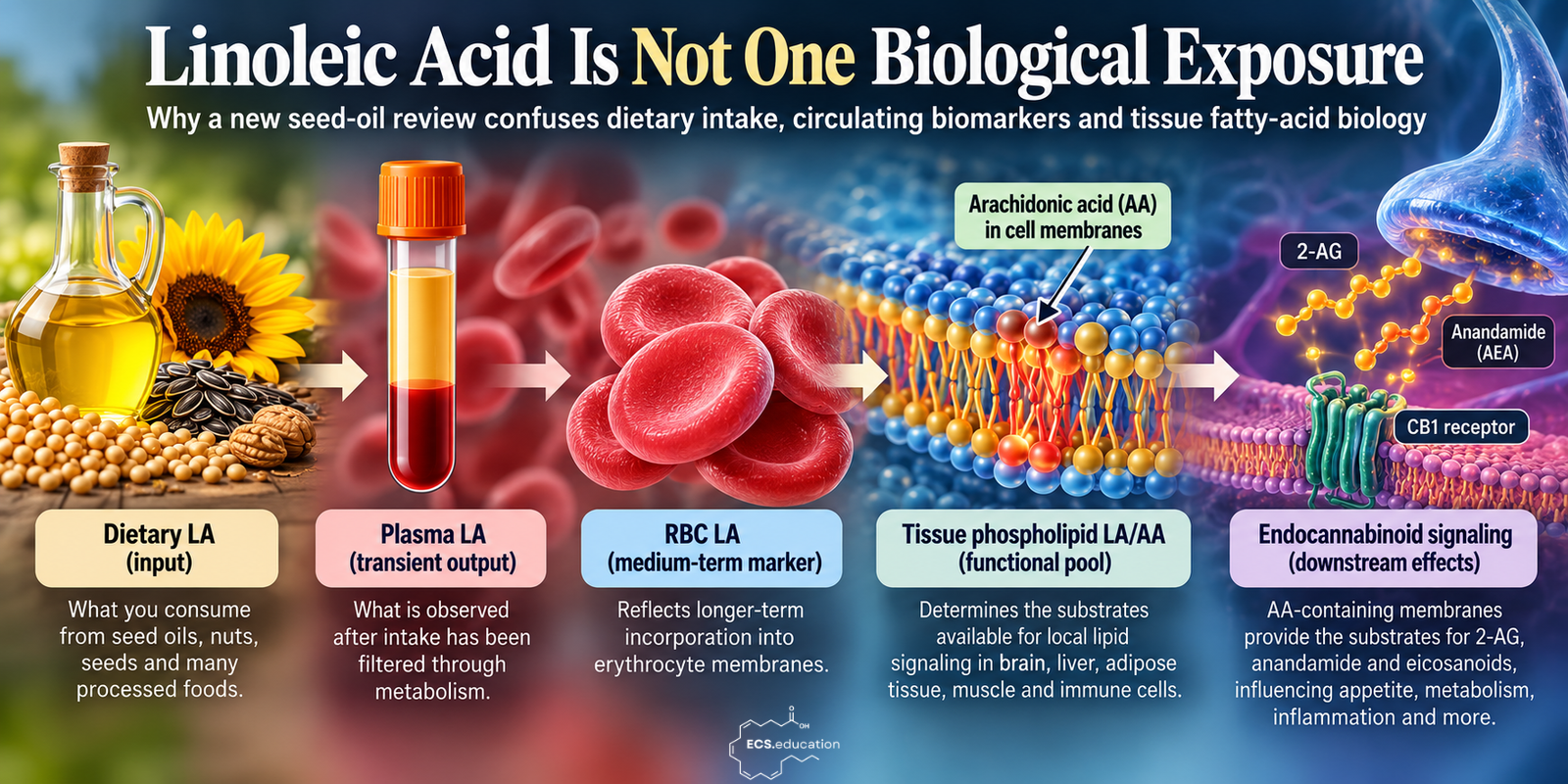
Dietary LA is an input: what you consume.
Plasma LA is an output, shaped by absorption, oxidation, hepatic lipid processing, desaturation, elongation, insulin sensitivity, and competition with other fatty acids.
RBC LA reflects long-term membrane incorporation.
Adipose LA represents storage.
Tissue phospholipid LA and arachidonic acid (AA) determine the actual substrates available for local lipid signaling.
These compartments are related, but they are not the same. Yet the review repeatedly shifts from:
Higher circulating LA → better outcome
to an implied conclusion:
Higher dietary LA → better outcome.
That leap is not justified.
Why Plasma Linoleic Acid Is Not the Same as Dietary Intake
This distinction is crucial because plasma LA can act as a marker of fatty-acid processing rather than simple intake.
LA follows the omega-6 pathway:
LA → GLA → DGLA → AA
with FADS2 and FADS1 controlling key desaturation steps.
Someone with lower downstream conversion may retain more LA upstream, while someone with higher desaturase activity may show lower LA alongside greater downstream omega-6 production—despite consuming the same amount of dietary LA.
This is why low plasma LA may sometimes signal high FADS activity rather than a need for more seed oil.
The new review largely overlooks this nuance. It even uses the phrase “circulating LA intake” when discussing MASLD (metabolic dysfunction-associated steatotic liver disease).
Circulating LA is not intake. It’s what remains after metabolism has acted on it.
Stable AA Does Not Mean Dietary LA Is Irrelevant
A recurring argument in the omega-6 literature is that increasing dietary LA does not substantially increase plasma or RBC arachidonic acid.
That observation is real. Rett and Whelan’s frequently cited systematic review found little evidence that altering LA intake within Western dietary ranges changes AA in plasma/serum phospholipids or erythrocytes (Rett & Whelan, 2011).
The interpretation commonly attached to that observation is the problem.
The logic becomes:
“Increasing LA does not increase RBC/plasma AA. Therefore dietary LA does not meaningfully determine tissue AA.”
That conclusion does not follow from those data.
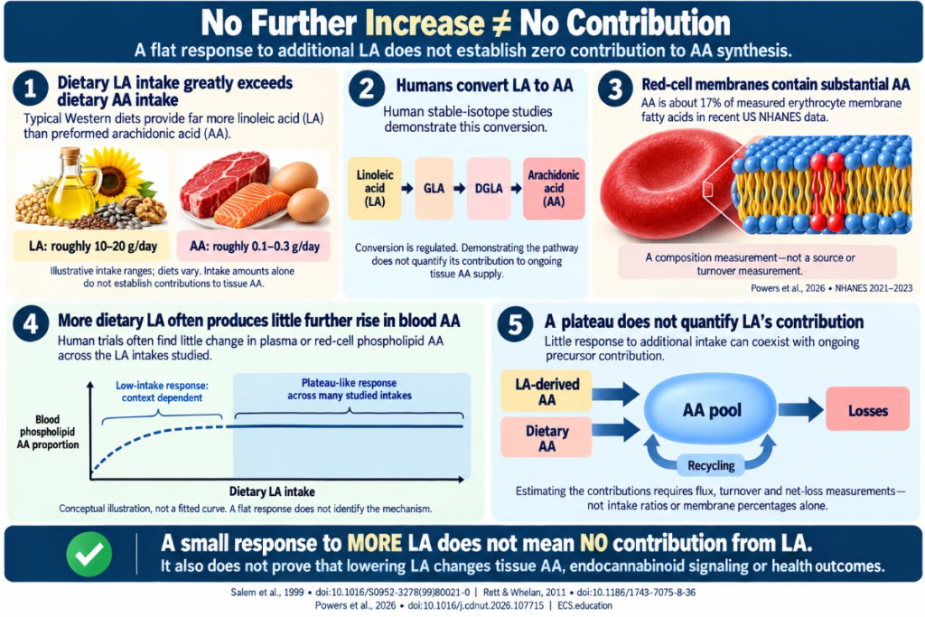
Humans demonstrably convert LA to AA in vivo. This has been shown directly using stable-isotope tracing (Salem et al., 1999).
Western diets also provide vastly more LA than preformed dietary AA. The substantial AA content of human membranes therefore cannot be explained by dietary AA alone.
A large part of that pool must ultimately be maintained through endogenous metabolism of dietary LA.
The key distinction is between precursor flux and steady-state pool size.
Once the downstream AA pool is established and tightly regulated, increasing LA further may do very little to raise it.
That is a plateau.
It is certainly not evidence of irrelevance.
No Further Increase Does Not Mean No Contribution
This is the core issue.
If the downstream AA pool is already near a homeostatically regulated plateau, increasing LA from one Western-level intake to another may produce little change in bulk RBC or plasma AA.
That tells us:
The AA pool is regulated.
It does not tell us:
LA did not help build or maintain that pool.
This distinction is simple but repeatedly ignored in the literature. It becomes even more critical when examining human membrane composition.
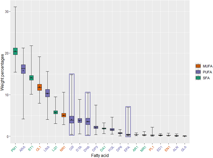
Recent nationally representative NHANES measurements show that arachidonic acid constitutes roughly 17% of measured fatty acids in American erythrocyte membranes, making it one of the most abundant fatty acids in the membrane, while EPA and DHA remain comparatively scarce (Powers et al., 2026).
That AA pool does not appear by chance. It is continuously maintained through fatty-acid metabolism and membrane remodeling.
The fact that additional dietary LA doesn’t push the pool higher doesn’t sever the biochemical link between LA and AA.
Blood Is Not Tissue Biology
Another problem is that much of the research used to argue that LA does not meaningfully alter AA biology relies on plasma, serum or erythrocyte measurements.
Rett and Whelan’s influential review is a useful example. Despite referring to “tissue arachidonic acid” in its title, the eligible human studies primarily measured plasma/serum phospholipids and red blood cell fatty acids (Rett & Whelan, 2011).
These are useful biomarkers.
But they’re not hepatocytes, adipocytes, hypothalamic neurons, astrocytes, skeletal muscle, or immune cells.
A stable RBC AA percentage doesn’t prove that arachidonoyl phospholipid availability is unchanged in metabolically relevant tissues. It tells us little about:
- Phospholipid class
- Molecular species
- Tissue turnover
- Local phospholipase activity
- DAG composition
- Omega-3 competition
- Downstream lipid mediator synthesis
This is where the endocannabinoid system (ECS) becomes impossible to ignore.
How Linoleic Acid Feeds Arachidonic Acid and the Endocannabinoid System
AA isn’t just a membrane fatty acid—it’s the backbone of the two primary endogenous CB1 agonists:
- 2-arachidonoylglycerol (2-AG)
- Anandamide (AEA)
The membrane isn’t passive storage—it’s a substrate reservoir for signaling.
When arachidonoyl-containing phospholipids are mobilized, they fuel the production of endocannabinoids, eicosanoids, and other bioactive lipids.
The real question isn’t:
What is the plasma AA concentration?
It’s:
What arachidonoyl substrates are available in the relevant tissue, and how are they being used?
The mechanistic evidence here is strong. Alvheim and colleagues showed experimentally that increasing dietary LA altered tissue arachidonoyl phospholipids and increased the AA-derived endocannabinoids 2-AG and anandamide. Adding EPA and DHA shifted the substrate pools and attenuated the phenotype (Alvheim et al., 2012).
Importantly, this is not solely an animal observation.
In a human dietary intervention, increasing omega-3 intake while lowering LA decreased the AA-derived endocannabinoid 2-AG by approximately 25%, while markedly increasing DHA-derived endocannabinoid-like mediators (Ramsden et al., 2015).
This is substrate-driven ECS biology.
And the concept is no longer peripheral to contemporary endocannabinoid science. A 2026 review of diet and the endocannabinoidome explicitly describes dietary fatty acids as regulators of this system through substrate availability, enzymatic competition and changes in bioactive lipid-mediator abundance (Petrosino et al., 2026).
None of this is captured adequately by CRP.
“LA Does Not Cause Inflammation” Answers the Wrong Question
The review emphasizes that clinical trials don’t show increased conventional inflammatory markers (e.g., CRP, TNF-α) with higher LA intake.
That may be true, but it doesn’t resolve the biological question.
CRP, TNF-α, and circulating cytokines are downstream inflammatory readouts. The real question is whether LA alters:
- Membrane arachidonate
- Endocannabinoid substrate availability
- Eicosanoid production
- Oxidized LA metabolites
- Receptor signaling
- Metabolic adaptation
A diet can reshape lipid signaling without causing a measurable rise in fasting CRP.
“CRP didn’t increase” does not mean “nothing biologically relevant changed.”
Why Mendelian Randomization of Circulating LA Is Not a Seed-Oil Trial
The skin-cancer section introduces another category error.
The review cites Mendelian-randomization studies where genetically predicted circulating LA is linked to lower risk of some skin cancers. This is then used to reassure readers about dietary LA.
But genetic variants tied to circulating LA often affect fatty-acid metabolism, particularly the FADS pathway.
This means the instrument may reflect:
How efficiently LA is converted downstream
rather than:
How much LA someone consumes.
A genetic tendency toward high circulating LA due to reduced conversion is biologically different from increasing dietary LA.
One is a metabolic phenotype, the other is a dietary intervention.
They should not be treated as interchangeable.
Replacement Trials Answer a Different Question
The liver-fat evidence also requires careful interpretation.
Several RCTs show that replacing saturated fat with PUFA reduces hepatic fat accumulation under specific conditions, including overfeeding.
This is meaningful evidence, but it answers:
“Is PUFA better than SFA in this experimental context?”
It does not answer:
“Is more LA always better?”
Nor does it establish that the fatty-acid composition of a modern Western diet is physiologically optimal.
Replacement experiments compare two interventions—they don’t define the biological optimum.
The Review’s Own Data Reveal the Missing Pathway
Some of the evidence cited by the authors actually points to the importance of downstream metabolism.
In skin-cancer research, genetically predicted LA and AA don’t behave the same way. Higher genetically predicted LA is sometimes linked to lower risk, while higher AA is linked to higher risk.
In another adipose-tissue study discussed in the review, AA differed between groups while LA did not.
This should raise an immediate question:
What happened between LA and AA?
That’s where desaturase activity, elongation, tissue remodeling, and substrate partitioning matter.
Instead, the pathway is repeatedly compressed into the label “LA exposure”.
We’ve Seen This Framing Before
This isn’t the first time an industry-linked review involving Mark Messina has discussed omega-6 while largely excluding tissue substrate biology and the endocannabinoid system. In our earlier analysis of omega-6 fats, seed oils and the endocannabinoid system, I dissected his earlier LA review using the same framing.
The pattern is familiar:
Circulating LA looks favorable → LA is interpreted as favorable → tissue AA and downstream signaling remain largely ignored.
That’s too simplistic.
Higher plasma LA isn’t the same as eating more LA.
Genetically elevated LA isn’t the same as consuming more soybean oil.
Stable RBC AA doesn’t mean dietary LA didn’t help generate the AA pool.
Normal CRP doesn’t prove a lack of altered lipid signaling.
Replacing saturated fat with PUFA doesn’t identify the optimal tissue fatty-acid composition.
The Right Question to Ask
The usual framing of the seed-oil debate is:
“Are seed oils good or bad?”
That’s the wrong question.
The biologically useful question is:
“What membrane and metabolic state does a given diet produce in a given person?”
To answer that, we need to follow the full chain:
Dietary input → metabolic conversion → tissue incorporation → membrane substrate architecture → lipid mediator production → receptor signaling → physiology
LA sits near the beginning of that chain.
Plasma LA sits somewhere in the middle.
Arachidonoyl phospholipids sit further downstream.
2-AG, anandamide, and eicosanoids emerge from that substrate architecture.
We can clearly see that these are not the same exposure.
Until nutrition science respects these distinctions, statistically strong associations with circulating LA will continue to tell us far less about tissue lipid biology than their headlines suggest.
Linoleic acid is not a single biological exposure.
And a blood concentration is not a substitute for understanding what the body did with the substrate.
References
Alvheim AR, Malde MK, Osei-Hyiaman D, et al. Dietary linoleic acid elevates endogenous 2-AG and anandamide and induces obesity. Obesity (Silver Spring). 2012;20(10):1984-1994. doi:10.1038/oby.2012.38
Messina M, Nagra M, Goldman D, Nechuta S. Evidence Suggests Linoleic Acid Exerts Health Benefits Beyond Cardiovascular Disease: A Brief Review of Clinical and Observational Studies. Curr Nutr Rep. 2026;15(1):87. Published 2026 Oct 22. doi:10.1007/s13668-026-00806-9
Petrosino S, Fontecilla-Escobar J, Di Marzo V, Iannotti FA. Diet, microbiota, and lipidomics: How fatty acids shape the endocannabinoidome and host metabolism. Prog Lipid Res. Published online July 7, 2026. doi:10.1016/j.plipres.2026.101396
Powers CD, Sternberg MR, Mineva EM, et al. Over half of the United States population had an undesirably low Omega-3 Index based on erythrocyte membrane measurements: results from the cross-sectional NHANES from August 2021 to August 2023. Curr Dev Nutr. 2026;10(6):107715. doi:10.1016/j.cdnut.2026.107715.
Ramsden CE, Zamora D, Makriyannis A, et al. Diet-induced changes in n-3- and n-6-derived endocannabinoids and reductions in headache pain and psychological distress. J Pain. 2015;16(8):707-716. doi:10.1016/j.jpain.2015.04.007
Rett BS, Whelan J. Increasing dietary linoleic acid does not increase tissue arachidonic acid content in adults consuming Western-type diets: a systematic review. Nutr Metab (Lond). 2011;8:36. Published 2011 Jun 10. doi:10.1186/1743-7075-8-36
Salem N Jr, Pawlosky R, Wegher B, Hibbeln J. In vivo conversion of linoleic acid to arachidonic acid in human adults. Prostaglandins Leukot Essent Fatty Acids. 1999;60(5-6):407-410. doi:10.1016/s0952-3278(99)80021-0