
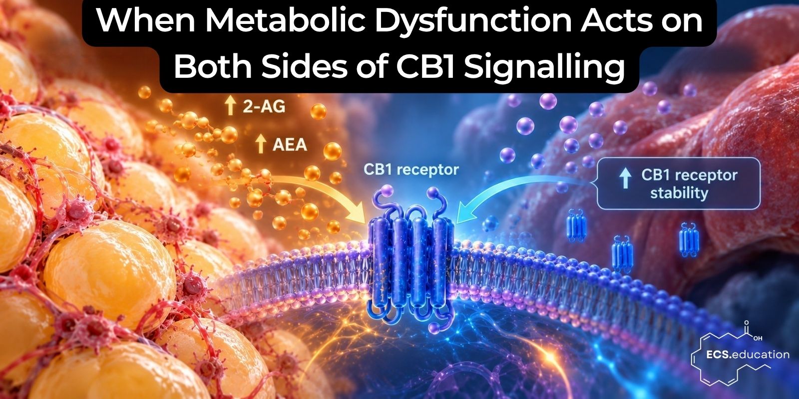
New human data on visceral fat strengthen the case for studying the endocannabinoid system as a metabolically regulated homeostatic system
Just over a month ago, we published an open letter asking whether metabolic state can regulate CB1 receptor stability across insulin-sensitive tissues. The immediate reason was a study by Liu and colleagues showing that insulin increases hepatic CB1 receptor protein through ERK-dependent suppression of lysosomal degradation, without requiring a corresponding increase in CNR1 transcription. The finding suggested that metabolic regulation of CB1 may occur not only through changes in ligand production, but also through changes in receptor turnover.
A new study published in Diabetologia now adds an important piece from the other side of the signalling equation.
Barbosa and colleagues examined endocannabinoid production directly in visceral and subcutaneous adipose tissue from mice and humans, including individuals with obesity with and without type 2 diabetes. Their central finding was not simply that visceral fat produces endocannabinoids. It was that endocannabinoid overproduction tracked metabolic dysfunction much more closely than adiposity itself.
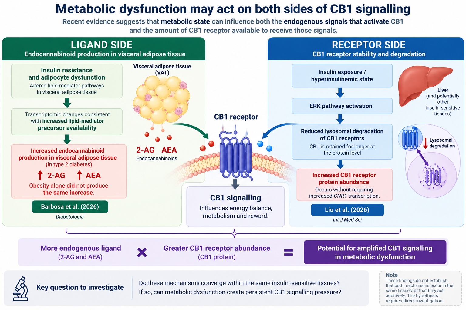
Human visceral adipose tissue produced approximately 1.9 times more 2-arachidonoylglycerol (2-AG) than subcutaneous adipose tissue. More importantly, visceral fat from individuals with obesity and type 2 diabetes showed approximately 1.7-fold greater 2-AG production and 1.8-fold greater anandamide (AEA) production compared with controls. Obesity in the absence of type 2 diabetes did not produce the same increase, and subcutaneous adipose tissue remained largely unaffected.
AEA production from visceral adipose tissue also correlated positively with HbA1c and fasting plasma glucose. This makes the metabolic phenotype particularly interesting. The increase in endocannabinoid production appears to follow deterioration in metabolic regulation rather than simply an increase in adipose mass.
Visceral adipose tissue changes as metabolic control deteriorates
Adipose tissue is not an inert energy store. It responds to insulin, regulates the uptake and release of metabolic substrates, secretes endocrine signals and lipid mediators, and communicates extensively with other tissues. It also possesses the enzymatic machinery required to synthesize and degrade endocannabinoids.
Barbosa and colleagues found that type 2 diabetes was associated with repression of insulin-sensitive adipocyte programmes together with activation of inflammatory and lipid-mediator pathways. Their cross-species transcriptomic analysis also identified changes consistent with increased availability of lipid precursors and increased purinergic receptor expression.
The relationship between these transcriptional changes and endocannabinoid production still needs to be resolved mechanistically, but the overall pattern is informative. Increased endocannabinoid production emerged in visceral adipose tissue as normal insulin-sensitive adipocyte biology was being lost.
This makes insulin resistance a more informative variable than obesity alone.
Two people can carry similar amounts of adipose tissue while differing substantially in insulin sensitivity, inflammatory status, adipocyte function, mitochondrial capacity and metabolic flexibility. The new data suggest that endocannabinoid production can differ with that physiological state as well.
Rather than thinking of obesity as a condition in which the ECS simply becomes more active, it may be more useful to consider whether metabolically dysfunctional tissues acquire a different ECS phenotype.
That distinction fits well with the broader argument of our recent open letter: the functional state of the ECS cannot be inferred from ligand concentrations alone.
The ligand side and the receptor side
Any receptor signalling system depends on both the amount of signal being generated and the state of the receptor population receiving that signal.
Barbosa et al. provide evidence for changes on the ligand side. In metabolically dysfunctional visceral adipose tissue, production of both 2-AG and AEA was increased.
Liu et al. provide evidence for a separate mechanism on the receptor side. In hepatocytes and liver, insulin activated ERK signalling and reduced lysosomal degradation of CB1, increasing CB1 protein abundance without requiring increased CNR1 transcription. Insulin pretreatment also enhanced the subsequent lipogenic response to CB1 receptor activation.
The studies were conducted in different tissues and do not demonstrate that these mechanisms occur together. Barbosa et al. did not investigate insulin-dependent stabilization of CB1 in adipocytes, and Liu et al. did not establish that their hepatic mechanism operates in visceral fat or other insulin-sensitive tissues.
They nevertheless identify two complementary mechanisms that can now be considered within the same framework:
increased production of endogenous CB1 ligands
together with
increased persistence of CB1 receptor protein
could increase the potential for CB1 signalling in metabolically dysfunctional tissue.
Whether those mechanisms actually converge within the same cells or tissues is now an obvious experimental question.
A potential metabolic feedback loop
The significance of this question extends beyond using CB1 as a marker of metabolic dysfunction. Peripheral CB1 signalling is itself involved in lipid handling, glucose regulation, energy storage and insulin sensitivity.
Work going back more than two decades has shown that hepatic CB1 activation can stimulate fatty-acid synthesis and contribute to diet-induced metabolic dysfunction. Subsequent studies have connected hepatic CB1 signalling to impaired insulin signalling and altered insulin clearance. Adipose CB1 signalling can likewise interfere with normal metabolic regulation.
This creates the possibility of a feedback loop rather than a simple downstream response.
As insulin sensitivity deteriorates, visceral adipocytes may increase their production of 2-AG and AEA. Greater endocannabinoid exposure can then increase CB1 signalling pressure in tissues where the receptor contributes to energy storage and impaired metabolic regulation. If hyperinsulinemia simultaneously increases CB1 protein persistence in some of those tissues, receptor abundance could provide an additional layer of amplification.
In simplified form, the sequence could look something like this:
insulin resistance and adipocyte dysfunction
↓
increased visceral 2-AG and AEA production
↓
increased CB1 signalling pressure
↓
altered lipid handling and insulin action
↓
further metabolic dysfunction
A second mechanism could operate in parallel:
insulin resistance
↓
compensatory hyperinsulinemia
↓
reduced CB1 degradation in susceptible tissues
↓
greater CB1 protein abundance
↓
greater signalling potential at a given ligand concentration
Neither sequence should presently be treated as an established whole-body mechanism. What has changed is that individual components of such a model are increasingly supported experimentally and can now be tested together.
Metabolic state may be more informative than “endocannabinoid tone”
This also highlights a limitation in the way the term “endocannabinoid tone” is sometimes used. It can give the impression that the ECS has a single global setting that is either high or low.
The biology is considerably more distributed.
Effective CB1 signalling depends on tissue-specific production of endocannabinoids, membrane lipid substrate availability, synthesis and degradation enzyme activity, receptor abundance, receptor localization, trafficking and turnover, coupling efficiency and the metabolic state of the cell.
Changes in any of these variables can alter the biological response to the same nominal concentration of ligand.
An individual could therefore have increased endocannabinoid production in visceral adipose tissue while showing very different CB1 regulation in liver, skeletal muscle or brain. Even within the same tissue, increased ligand production does not tell us whether receptors are abundant, desensitized, internalized, efficiently coupled or undergoing altered turnover.
This is why describing complex chronic disease as simply a state of “endocannabinoid deficiency” or “endocannabinoid excess” is unlikely to capture the relevant physiology.
The more useful question is what state the ECS is operating in, in which tissue, and under what metabolic conditions.
Diet may regulate the ECS at several levels
The Barbosa study also strengthens a broader idea raised in our open letter: that the ECS may need to be understood as a diet-responsive adaptive homeostatic system.
This does not mean that a particular food or macronutrient directly determines ECS activity in a simple linear way. Rather, nutritional exposure can potentially affect several components of the signalling architecture.
The first is the membrane substrate environment. Endocannabinoids are lipid-derived signalling molecules, and the fatty-acid composition of cellular membranes influences the lipid pools from which those signals are generated.
The second is regulation of endocannabinoid synthesis and degradation. Barbosa et al. now show that insulin-resistant visceral adipose tissue can enter a state of increased 2-AG and AEA production.
The third is hormonal state. Insulin conveys information about nutrient availability, and chronic hyperinsulinemia is a characteristic feature of many insulin-resistant states.
The fourth is receptor regulation. Liu et al. demonstrate, at least in liver, that insulin can influence how rapidly CB1 protein is degraded.
These mechanisms are distinct, but they need not be biologically independent. Dietary fat quality may alter the substrate environment from which lipid mediators are generated, while carbohydrate exposure and the resulting insulin dynamics influence the hormonal environment in which those pathways operate. Once insulin resistance develops, regulation of both endocannabinoid production and potentially receptor turnover may change further.
This is a considerably broader model of ECS regulation than the one obtained by viewing the system primarily through exogenous cannabinoid pharmacology.
The next experiment has become clearer
The Barbosa paper points directly to one particularly relevant experiment:
Does insulin regulate CB1 receptor stability in visceral adipocytes, and does insulin resistance change that relationship?
An informative experiment would compare insulin-sensitive and insulin-resistant adipocytes while measuring CB1 protein abundance, CNR1 transcript, ERK activation, receptor turnover, lysosomal localization, internalization and recycling, endocannabinoid production and functional downstream CB1 signalling.
If insulin increases CB1 protein without a corresponding increase in CNR1 transcript, and if the effect is accompanied by altered lysosomal trafficking, that would provide evidence that the hepatic mechanism described by Liu extends into adipose tissue.
The experiment would become even more informative if performed alongside measurements of 2-AG and AEA production, because it would allow ligand-side and receptor-side regulation to be assessed in the same cellular system.
The same logic could subsequently be applied to other insulin-responsive tissues, including skeletal muscle, cardiac tissue, pancreatic islets and selected neuronal populations. There is no reason to assume that CB1 will be regulated identically in all of them. Establishing where the mechanism operates, where it does not, and how insulin resistance changes it would itself be a useful map of metabolic CB1 biology.
Brain regions such as the hypothalamus, hippocampus, striatum and nucleus accumbens are particularly interesting because they sit at the intersection of metabolic signalling, feeding, reward, stress and behavioural adaptation. Our open letter was partly motivated by the observation that short-term excessive sucrose exposure increased hippocampal CB1 binding in Göttingen minipigs. That finding does not establish an insulin-mediated mechanism, but it does suggest that central CB1 availability can respond to dietary metabolic conditions on relatively short timescales.
Metabolic dysfunction may remodel the ECS itself
The most interesting conclusion from Barbosa et al. is therefore not simply that people with type 2 diabetes have increased endocannabinoid production in visceral fat.
It is that the ECS appears to change as the metabolic state of the tissue deteriorates.
Placed beside the Liu findings, this suggests that metabolic regulation may operate on both the endogenous signals entering the CB1 system and the receptor population available to receive those signals.
We do not yet know whether the two mechanisms converge within the same insulin-sensitive tissues, whether their effects are additive, or whether compensatory mechanisms eventually oppose them. Those questions require direct experimental work.
Importantly, the emerging model is testable.
Metabolic dysfunction may not merely produce downstream consequences that happen to involve the ECS. It may actively remodel components of the ECS itself, potentially creating feedback between insulin resistance, lipid signalling, receptor regulation and energy metabolism.
If that is correct, the implications extend beyond obesity and type 2 diabetes. They would support treating the ECS as part of mainstream metabolic physiology rather than as a specialised signalling system primarily relevant to cannabis pharmacology.
CB1 activation is not inherently pathological, just as insulin signalling, inflammatory signalling or sympathetic activation are not inherently pathological. These systems become problematic when their magnitude, duration or tissue context no longer matches physiological demand.
A homeostatic signalling system that normally helps organisms adapt to changes in nutrient availability can likewise become maladaptive when the metabolic environment driving it remains chronically abnormal.
Understanding when that transition occurs, and whether it can be reversed, is now a much more interesting question than asking whether the ECS is simply “high” or “low”.
References
- Barbosa R, Leemput J, Passilly-Degrace P, et al. Visceral fat endocannabinoid overproduction is associated with insulin resistance and tissue dysfunction in mice and humans. Diabetologia. Published online October 5, 2026. doi:10.1007/s00125-026-06886-0.
- Liu SY, Ting CH, Hwang GS, et al. Insulin increases expression of cannabinoid receptor 1 by suppressing lysosomal degradation via ERK signaling pathway. Int J Med Sci. 2026;23(6):2027-2038. doi:10.7150/ijms.126308.
- Liu J, Zhou L, Xiong K, et al. Hepatic cannabinoid receptor-1 mediates diet-induced insulin resistance via inhibition of insulin signaling and clearance in mice. Gastroenterology. 2012;142(5):1218-1228.e1. doi:10.1053/j.gastro.2012.01.032.
- Osei-Hyiaman D, DePetrillo M, Pacher P, et al. Endocannabinoid activation at hepatic CB1 receptors stimulates fatty acid synthesis and contributes to diet-induced obesity. J Clin Invest. 2005;115(5):1298-1305. doi:10.1172/JCI23057.
- Muller T, Demizieux L, Troy-Fioramonti S, et al. Overactivation of the endocannabinoid system alters the antilipolytic action of insulin in mouse adipose tissue. Am J Physiol Endocrinol Metab. 2017;313(1). doi:10.1152/ajpendo.00374.2016.